43 easy microscope diagram with labels
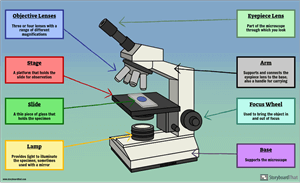
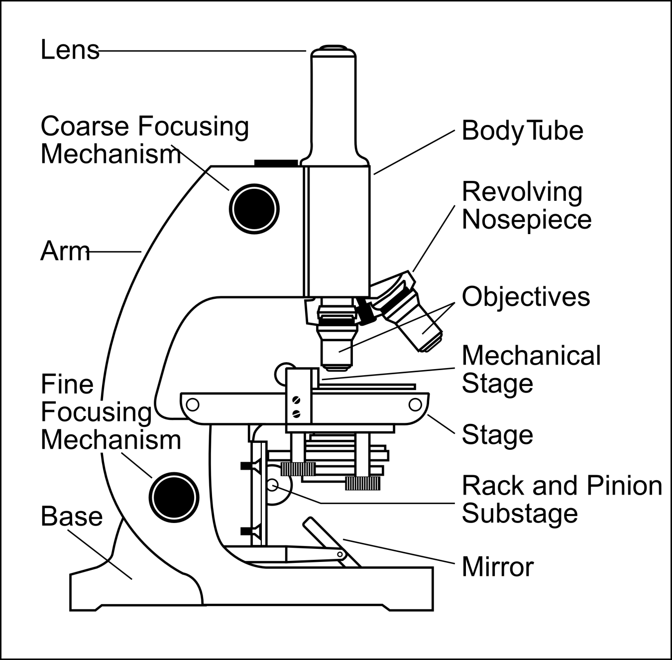
Label Microscope Diagram - EnchantedLearning.com arm - this attaches the eyepiece and body tube to the base. base - this supports the microscope. body tube - the tube that supports the eyepiece. coarse focus adjustment - a knob that makes large adjustments to the focus. diaphragm - an adjustable opening under the stage, allowing different amounts of light onto the stage. Types of Microscopes: Definition, Working Principle, Diagram ... Where, D is the least distinct vision; F is the focal length of the convex lens; Simple Microscope Diagram. Principle of Simple Microscope. The working principle of a simple microscope is that when a sample is placed within the focus of the microscope, a virtual, erect and magnified image is obtained at the least distance of distinct vision from the eye that is held at the lens.
Free Microscope Worksheets for Simple Science Fun for Your Students 1. Parts of a Microscope . The first worksheet labels the different parts of a microscope, including the base, slide holder, and condenser. If you have a microscope, compare and contrast this worksheet to it.Also, your kids can color this microscope diagram in and read the words to each part of the microscope.
Easy microscope diagram with labels
Label the microscope — Science Learning Hub Label the microscope Add to collection Use this interactive to identify and label the main parts of a microscope. Drag and drop the text labels onto the microscope diagram. eye piece lens coarse focus adjustment high-power objective diaphragm or iris base fine focus adjustment light source stage Download Exercise Tweet Microscope, Microscope Parts, Labeled Diagram, and Functions Revolving Nosepiece or Turret: Turret is the part of the microscope that holds two or multiple objective lenses and helps to rotate objective lenses and also helps to easily change power. Objective Lenses: Three are 3 or 4 objective lenses on a microscope. The objective lenses almost always consist of 4x, 10x, 40x and 100x powers. The most common eyepiece lens is 10x and when it coupled with ... Microscope labeled diagram - SlideShare Microscope labeled diagram 1. The Microscope Image courtesy of: Microscopehelp.com Basic rules to using the microscope 1. You should always carry a microscope with two hands, one on the arm and the other under the base. 2. You should always start on the lowest power objective lens and should always leave the microscope on the low power lens ...
Easy microscope diagram with labels. PDF Parts of a Microscope Printables - Homeschool Creations Label the parts of the microscope. You can use the word bank below to fill in the blanks or cut and paste the words at the bottom. Microscope Created by Jolanthe @ HomeschoolCreations.net. Parts of a eyepiece arm stageclips nosepiece focusing knobs illuminator stage objective lenses Simple Squamous Epithelium under a Microscope with a Labeled Diagram ... Simple columnar epithelium labeled. This is a labeled diagram of a simple columnar epithelium under a light microscope. I tried to show you both ciliated and nonciliated simple columnar epithelium. These diagrams show the cilia on the cell surface, rectangular cell, and elongated nucleus. Simple Microscope - Definition, Types, Working Principle & Formula 1. Simple microscope comprises a biconvex lens used as a magnifying glass. Compound microscope comprises 2 or more convex lenses where one lens is the eyepiece and the other one is the objective lens. 2. Natural light is the source to see the object. An illuminator is a source to see the object. 3. Parts of a microscope with functions and labeled diagram Microscope compound drawing light simple labeled cartoon easy openstax van clipart cliparts library parts grade dele jobilize mirror cnx complete . Description. Parts of a microscope with functions and labeled diagram is free image that you can download for free in My Awesome Site. This Parts of a microscope with functions and labeled diagram ...
Microscope Poster - Diagram with Labels | Teach Starter A poster containing a diagram with labels showing the key parts of a microscope. Use this educational classroom poster in your science lessons to highlight the key parts of a microscope. A lot of equipment is used in science experiments and it is important to know the names of and understand each part of the equipment and how it works. Simple Microscope - Diagram (Parts labelled), Principle, Formula and Uses Parts of a Simple Microscope A simple microscope consists of Optical parts Mechanical parts Labeled Diagram of simple microscope parts Optical parts The optical parts of a simple microscope include Lens Mirror Eyepiece Lens A simple microscope uses biconvex lens to magnify the image of a specimen under focus. Microscope Types (with labeled diagrams) and Functions Electron microscope labeled diagram The different types of electron microscopes are: Transmission Electron Microscope Scanning Electron Microscope Reflection Electron Microscope Scanning transmission electron microscope Scanning tunneling microscopy Electron microscope functions: Semiconductors and Data Storage Industry Failure Analysis Microscope Labeling - The Biology Corner Students label the parts of the microscope in this photo of a basic laboratory light microscope. Can be used for practice or as a quiz. ... The type of microscope used in most science classes is the _____ microscope. 18. You should carry the microscope by the _____ and the _____. 19. The objectives are attached to what part of the microscope ...
Parts of the Microscope with Labeling (also Free Printouts) Parts of the Microscope with Labeling (also Free Printouts) A microscope is one of the invaluable tools in the laboratory setting. It is used to observe things that cannot be seen by the naked eye. Table of Contents 1. Eyepiece 2. Body tube/Head 3. Turret/Nose piece 4. Objective lenses 5. Knobs (fine and coarse) 6. Stage and stage clips 7. Aperture 16 Parts of a Compound Microscope: Diagrams and Video Once you have an understanding of the parts of the microscope it will be much easier to navigate around and begin observing your specimen, which is the fun part! The 16 core parts of a compound microscope are: Head (Body) Arm. Base. Eyepiece. Eyepiece tube. Simple Microscope - Parts, Functions, Diagram and Labelling Picture 1: The image above is a stereo microscope. Image source: made-in-china.com Picture 2: The image above is a confocal microscope. Image source:thorlabs.com Picture 3: The image above is parts of scanning electron microscope. Image source:britannica.com Picture 4: The picture is a transmission electron microscope. Image source: ysjournal.com Pin on Science worksheets - Pinterest Download Clker's Microscope With Labels clip art and related images now. Multiple sizes and related images are all free on Clker.com. A diagram showing all of the parts of a compound light microscope. Mar 7, 2021 - This Pin was discovered by Gloria Legg. Discover (and save!) your own Pins on Pinterest.
Parts of a Microscope Labeling Activity
Compound Microscope Parts - Labeled Diagram and their Functions - Rs ... The eyepiece (or ocular lens) is the lens part at the top of a microscope that the viewer looks through. The standard eyepiece has a magnification of 10x. You may exchange with an optional eyepiece ranging from 5x - 30x. [In this figure] The structure inside an eyepiece. The current design of the eyepiece is no longer a single convex lens.

Easy labeled diagram of Microscope - YouTube
Label a Microscope - Storyboard That Create a poster that labels the parts of a microscope and includes descriptions of what each part does. Click "Start Assignment". Use a landscape poster layout (large or small). Search for a diagram of a microscope. Using arrows and textables label each part of the microscope and describe its function. Copy This Storyboard* More options

33 Microscope Label And Functions - Labels For You
Labeling the Parts of the Microscope | Microscope World Resources Labeling the Parts of the Microscope This activity has been designed for use in homes and schools. Each microscope layout (both blank and the version with answers) are available as PDF downloads. You can view a more in-depth review of each part of the microscope here. Download the Label the Parts of the Microscope PDF printable version here.
Compound Light Microscope Drawing at GetDrawings | Free download
Microscope Labeling - The Biology Corner 1) Start with scanning (the shortest objective) and only use the COARSE knob . Once it is focused… 2) Switch to low power (medium) and only use the COARSE knob . You may need to recenter your slide. Once it is focused.. 3) Switch to high power (long objective).

32 Label The Structures Of A Skeletal Muscle Fiber - Label Design Ideas 2020
Microscope Drawing | How To Draw A Microscope Diagram | Easy And Simple ... How to draw a microscope diagram. Microscope drawing. Easy and simple step by step tutorial for beginners.Thanks for Watching and Subscribing "Minutes Draw"M...

Microscope Labelled Diagram Gcse - Micropedia
Compound Microscope Parts, Functions, and Labeled Diagram Eyepiece (ocular lens) with or without Pointer: The part that is looked through at the top of the compound microscope. Eyepieces typically have a magnification between 5x & 30x. Monocular or Binocular Head: Structural support that holds & connects the eyepieces to the objective lenses. Arm: Supports the microscope head and attaches it to the base.
Structure and Function of Plants (Lab Practical #1) Flashcards | Easy Notecards
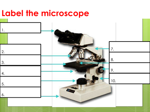
Parts Of The Microscope Label Worksheets & Teaching Resources | TpT Check out this simple and clear 13 question worksheet that covers the parts of a microscope. Their is a visual at the top of the page that points to 13 parts. ... This simple 1-page worksheet has students label a diagram of a microscope using words from a word bank. Subjects: Basic Principles, Biology, General Science. Grades: 7 th - 12 th ...

Male Reproductive System | Free Images at Clker.com - vector clip art online, royalty free ...
Parts of a Simple Microscope - Labeled (with diagrams) image 1: The images above are all examples of a simple microscope. image source: laboratoryinfo.com image 2: A simple microscope commonly used by students for studying minute objects. image source: imimg.com picture 3: It is the latest design of a simple microscope - advanced features than the conventional simple microscopes.

Oncology Basics 2016: Understanding the Cells
Microscope Drawing Easy with Label - YouTube In this video I go over a microscope drawing that is easy with label. There is a blank copy at the end of the video to review on your own. A great way to s...

ปักพินโดย ð“¥ð“®ð“𓪠ð“. ใน Biology | Science biology Secondary school science และ Science diagrams
Labelled Diagram of Compound Microscope - Biology Discussion The below mentioned article provides a labelled diagram of compound microscope. Part # 1. The Stand: The stand is made up of a heavy foot which carries a curved inclinable limb or arm bearing the body tube. The foot is generally horse shoe-shaped structure (Fig. 2) which rests on table top or any other surface on which the microscope in kept.
Microscope Labelled Diagram Gcse - Micropedia
Microscope labeled diagram - SlideShare Microscope labeled diagram 1. The Microscope Image courtesy of: Microscopehelp.com Basic rules to using the microscope 1. You should always carry a microscope with two hands, one on the arm and the other under the base. 2. You should always start on the lowest power objective lens and should always leave the microscope on the low power lens ...

Introductory Biology
Microscope, Microscope Parts, Labeled Diagram, and Functions Revolving Nosepiece or Turret: Turret is the part of the microscope that holds two or multiple objective lenses and helps to rotate objective lenses and also helps to easily change power. Objective Lenses: Three are 3 or 4 objective lenses on a microscope. The objective lenses almost always consist of 4x, 10x, 40x and 100x powers. The most common eyepiece lens is 10x and when it coupled with ...

A Study of the Microscope and its Functions With a Labeled Diagram | Diagrams/Charts/Maps ...
Label the microscope — Science Learning Hub Label the microscope Add to collection Use this interactive to identify and label the main parts of a microscope. Drag and drop the text labels onto the microscope diagram. eye piece lens coarse focus adjustment high-power objective diaphragm or iris base fine focus adjustment light source stage Download Exercise Tweet

Microscope 8 | Coloring pages, Coloring pages for kids, Printable coloring pages
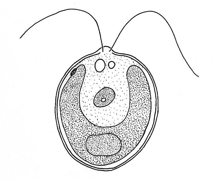
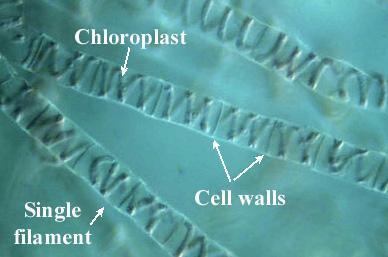
Chlamydomonas Diagram With Labels

All Saints Online: Diagram for Labelling: Microscope

All Saints Online: Diagram for Labelling: Microscope
Post a Comment for "43 easy microscope diagram with labels"